Anti-CD3 activation epitope Monoclonal Antibody (Clone:APA1/1)
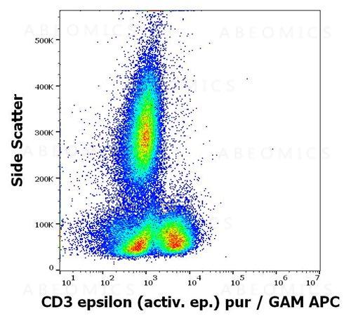
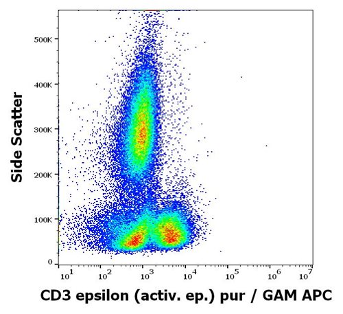
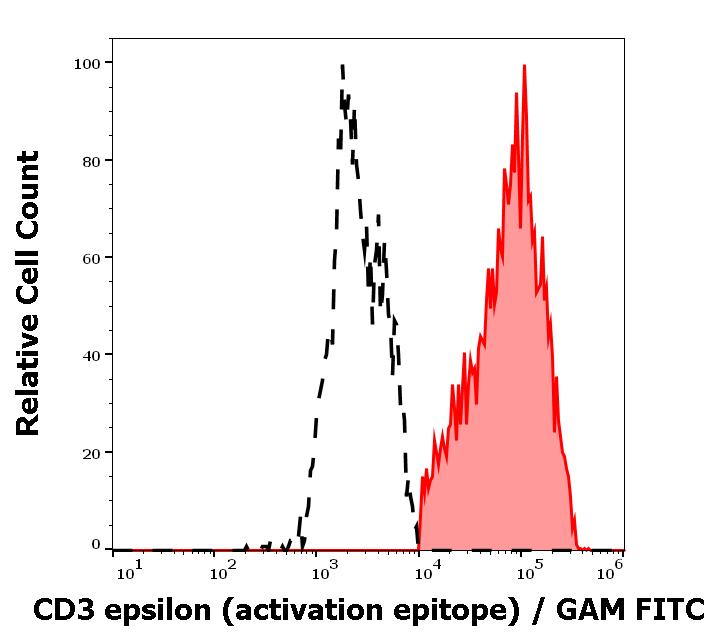
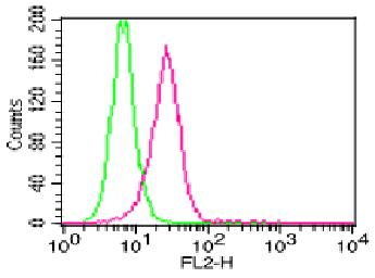
Figure 1: Flow cytometry intracellular staining pattern of PMA+Ionomycin stimulated human peripheral whole blood stained using anti-human CD3 epsilon activation epitope (APA1/1) purified antibody (concentration in sample 5 μg/ml, GAM APC).
Roll over image to zoom in
Shipping Info:
For estimated delivery dates, please contact us at [email protected]
| Format : | Purified |
| Amount : | 0.1 mg |
| Isotype : | Mouse IgG1 |
| Purification : | Purified by protein-A affinity chromatography |
| Storage condition : | Store at 2-8°C. Do not freeze. |
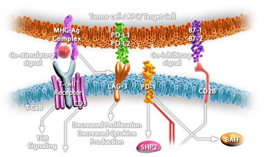
CD3 complex is crucial in transducing antigen-recognition signals into the cytoplasm of T cells and in regulating the cell surface expression of the TCR complex. T cell activation through the antigen receptor (TCR) involves the cytoplasmic tails of the CD3 subunits CD3 gamma, CD3 delta, CD3 epsilon and CD3 zeta. These CD3 subunits are structurally related members of the immunoglobulins super family encoded by closely linked genes on human chromosome 11. The CD3 components have long cytoplasmic tails that associate with cytoplasmic signal transduction molecules. This association is mediated at least in part by a double tyrosine-based motif present in a single copy in the CD3 subunits. CD3 may play a role in TCR-induced growth arrest, cell survival and proliferation.
Flow Cytometry Positive control: Human T cells stimulated with anti-CD3 (MEM-57) antibody (1 µg/ml)
Sample preparation:
At the end of stimulation of T cells, perform staining of surface markers (if required) in PBS + 0.1% BSA for 20 min. on ice.
Wash with PBS and fix with 2% formaldehyde, 30 min on ice.
Wash with PBS and incubate in PBS + 0,1% saponine, 5 min. RT.
Incubate the cells in PBS + 1% BSA + 0.03% saponine, 15 min. on ice.
Incubate with fluorescence-labeled APA1/1 antibody (1-2 µg/ml) in PBS + 1% BSA + 0.03% saponine in dark, 20 min. RT.
Wash with PBS + 1% BSA + 0.03% saponine, resuspend in PBS.
Immunoprecipitation Western Blotting Immunohistochemistry (frozen sections) Immunocytochemistry Application note:Fixed and permeabilised cells. The antibody can distinguish TCR-stimulated from non-stimulated cells.
For Research Use Only. Not for use in diagnostic/therapeutics procedures.
| Subcellular location: | Cell membrane |
| Post transnational modification: | Phosphorylated on Tyr residues after T-cell receptor triggering by LCK in association with CD4/CD8. |
| Tissue Specificity: | Highly expressed in T-helper cells. The presence of CD4 is a hallmark of T-helper cells which are specialized in the activation and growth of cytotoxic T-cells, regulation of B cells, or activation of phagocytes. CD4 is also present in other immune cells such as macrophages, dendritic cells or NK cells. |
| BioGrid: | 107354. 25 interactions. |
|
There are currently no product reviews
|



.png "ACE2/HEK293 Stable Cell Line")