Monoclonal Antibody to Dc-Sign/CD 209 (Clone: ABM47B7)

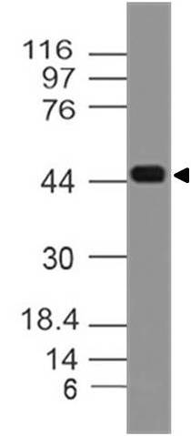
Fig-1: Expression analysis of Dc-Sign. Anti-Dc-Sign antibody (Clone: ABM47B7) was tested at 0.1 µg/ml on partial length recombinant protein.
Roll over image to zoom in
Shipping Info:
Order now and get it on Tuesday February 24, 2026
Same day delivery FREE on San Diego area orders placed by 1.00 PM
| Format : | Purified |
| Amount : | 100 µg |
| Isotype : | Mouse IgG2b Kappa |
| Purification : | Protein G Chromatography |
| Content : | 25 µg in 50 µl/100 µg in 200 µl PBS containing 0.05% BSA and 0.05% sodium azide. Sodium azide is highly toxic. |
| Storage condition : | Store the antibody at 4°C; stable for 6 months. For long-term storage; store at -20°C. Avoid repeated freeze and thaw cycles. |
Dendritic cell-specific intercellular adhesion molecule-3-grabbing non-integrin (DC-SIGN) is a tetrameric C-type (calcium-dependent) lectin that binds, through its C-terminal carbohydrate recognition domain, high mannose N-linked glycans present on the surface of several viral glycoproteins such as human immunodeficiency virus (HIV) gp120 and hepatitis C virus (HCV) E2. It facilitates DC-specific delivery of Ag. This is accomplished by conjugating Ag to receptor-specific Ab or carbohydrate ligands that bind to its carbohydrate recognition domain. In humans, DC-SIGN expression is restricted to DCs and certain types of macrophages. DC-SIGN is involved in the innate immune system and recognizes numerous evolutionarily divergent pathogens, including viruses, bacteria, fungi, and parasites. After binding, these pathogens are internalized and pathogen-derived antigens are presented via MHC class I and II molecules to CD8+ and CD4+ T cells, respectively. DC-SIGN represents a promising CLR for targeted vaccine delivery.
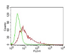
Western blot analysis: 0.1-0.5 µg/ml; FACS: 0.5-1 µg/10^6 Cells
Reference for expression of Dc-Sign:
Wai K. Lai, Phoebe J. Sun, Jie Zhang, Adam Jennings, Patricia F. Lalor, Stefan Hubscher,Jane A. McKeating,and David H. Adams. Expression of DC-SIGN and DC-SIGNR on Human Sinusoidal Endothelium Am J Pathol. 2006 Jul; 169(1): 200–208.doi: 10.2353/ajpath.2006.051191
For Research Use Only. Not for use in diagnostic/therapeutics procedures.
| Subcellular location: | Secreted |
| Tissue Specificity: | Predominantly expressed in dendritic cells and in DC-residing tissues. Also found in placental macrophages, endothelial cells of placental vascular channels, peripheral blood mononuclear cells, and THP-1 monocytes. |
| BioGrid: | 119051. 14 interactions. |
|
There are currently no product reviews
|


.png)


