

Monoclonal Antibody to Laminin, gamma 1 (LAMC1)(Clone : A5) (discontinued)
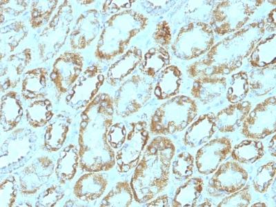
Formalin-fixed, paraffin-embedded human Renal Cell Carcinoma stained with Laminin Monoclonal Antibody (A5).
(Clone : A5) (discontinued)")
Roll over image to zoom in
Shipping Info:
For estimated delivery dates, please contact us at [email protected]
| Format : | Purified |
| Amount : | 100 µg |
| Isotype : | Rat IgG2a, kappa |
| Purification : | Affinity Chromatography |
| Content : | 100 µg in 500 µl PBS containing 0.05% BSA and 0.05% sodium azide. Sodium azide is highly toxic. |
| Storage condition : | Store the antibody at 4°C; stable for 6 months. For long-term storage; store at -20°C. Avoid repeated freeze and thaw cycles. |
| Gene : | Lamc1 |
| Uniprot ID : | P02468 |
| Alternative Name : | LAMC1, LAMB2 |
| Immunogen Information : | Murine EHS laminin prepaRation |
Laminins are large hetero-trimeric, non-collagenous glycoproteins composed of alpha , beta , and gamma chains. This MAb reacts with laminin B2/1 chain of ~210kDa and does not cross-react with other basement membrane components or fibronectin. Its specificity was established by immunoprecipitation and immunofluorescence of human skeletal muscle and kidney with laminin chain-specific MAbs. Epithelial sheets in vivo are separated from the mesenchymal elements of the stroma by a thin layer of a specialized type of extracellular matrix termed the basement membrane (BM). This structure consists of individual components, some of which are ubiquitous in BMs and some are not. The ubiquitous ones comprise laminin (LN), entactin/nidogen (EN), collagen type IV (CIV), and large heparan sulfate proteoglycan (HSPG), which interact specifically with each other to form a continuous and regular BM. Alterations of BM integrity, from local discontinuities up to complete loss, are described in many types of human and animal epithelial neoplasms. This MAb stains uniformly all human and murine basement membranes.
Flow Cytometry (0.5-1ug/million cells); Immunofluorescence (0.5-1.0ug/ml); Immunohistochemistry (Formalin-fixed) (1-2ug/ml for 30 minutes at RT)(Staining of formalin-fixed tissues requires heating tissue sections in 10mM Tris with 1mM EDTA, pH 9.0, for 45 min at 95 °C followed by cooling at RT for 20 minutes)
For Research Use Only. Not for use in diagnostic/therapeutics procedures.
| Subcellular location: | Secreted |
| Tissue Specificity: | Found in the basement membranes (major component). |
| BioGrid: | 110109. 35 interactions. |
|
There are currently no product reviews
|
 (Discontinued)")

; IgG1 Chimeric mAb")
")

 Monoclonal Antibody (Clone: TM228)(Discontinued)")
(KRTL/1077 + KRTH/1076)")
(Clone : MUC6/916)")
 (Mitochondrial Marker)(Clone : GROEL/730)")
(Clone : M2-9E3)")
(Clone : SPM588)")
(Clone : PR501)")
(Clone : A103)")
 (Clone: ABM1C12)")


")
")
.png "ACE2/HEK293 Stable Cell Line")
")


(Clone : SPM125)")
(Clone : 3E2D10)")
(Clone : GP1.4)")


