

Monoclonal antibody to Mouse CD4 (Clone: GK1.5) FITC Conjugated
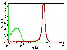
Fig:1-Cell surface flow analysis of mCD4 FITC cojugated in Mouse thymocytes using 0.5 µg/10^6 cells of mCD4 antibody (Clone: GK1.5). Green represents isotype control; red represents anti-mCD4 FITC antibody (10-4092F).
 FITC Conjugated")
Roll over image to zoom in
Shipping Info:
Order now and get it on Tuesday July 14, 2026
Same day delivery FREE on San Diego area orders placed by 1.00 PM
| Format : | Purified |
| Amount : | 100 µg |
| Isotype : | Rat IgG2b Kappa |
| Purification : | Protein G Chromatography |
| Content : | 0.2 mg/ml in Tris buffer containing 0.05% Azide |
| Storage condition : | Store the antibody at 4°C; stable for 6 months. |
FACS Analysis: 0.5-1 µg/10^6 cells
For Research Use Only. Not for use in diagnostic/therapeutics procedures.
| Subcellular location: | Cell membrane |
| Post transnational modification: | Phosphorylated by PKC; phosphorylation plays an important role for CD4 internalization. |
| Tissue Specificity: | Highly expressed in T-helper cells. The presence of CD4 is a hallmark of T-helper cells which are specialized in the activation and growth of cytotoxic T-cells, regulation of B cells, or activation of phagocytes. CD4 is also present in other immune cells such as macrophages, dendritic cells or NK cells. |
| BioGrid: | 198599. 1 interactions. |
|
There are currently no product reviews
|
(Discontinued)")

")
")

")
(Discontinued)")
(Discontinued)")
(Discontinued)")
 (Clone: ABM1C12)")


")
")
.png "ACE2/HEK293 Stable Cell Line")
")


")


