

Monoclonal Antibody to Cytokeratin, pan (Epithelial Marker)(Clone : AE-1/AE-3)
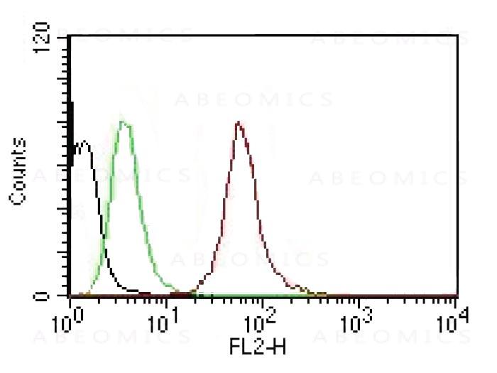
Figure 1: Flow Cytometric analysis of human Pan Cytokeratin on HeLa cells. Black: cells alone; Green: Isotype Control; Red: PE-labeled Pan-Cytokeratin Monoclonal Antibody (AE-1/AE-3).
(Clone : AE-1/AE-3)")
(Clone : AE-1/AE-3)")
Roll over image to zoom in
Shipping Info:
For estimated delivery dates, please contact us at [email protected]
| Format : | Purified |
| Amount : | 100 µg |
| Isotype : | Mouse IgG1 / Kappa |
| Purification : | 200ug/ml of Ab Purified from Bioreactor Concentrate by Protein A/G. |
| Content : | Prepared in 10mM PBS with 0.05% BSA & 0.05% azide. Also available WITHOUT BSA & azide at 1.0mg/ml. |
| Storage condition : | Store the antibody at -20 to -80°C. Antibody is stable for 24 months. Nonhazardous. No MSDS required. |
Twenty human keratins are resolved with two-dimensional gel electrophoresis into acidic (pI 6.0) subfamilies. This antibody cocktail recognizes acidic (Type I or LMW) and basic (Type II or HMW) cytokeratins, which 67kDa (CK1); 64kDa (CK3); 59kDa (CK4); 58kDa (CK5); 56kDa (CK6); 52kDa (CK8); 56.5kDa (CK10); 50kDa (CK14); 50kDa (CK15); 48kDa (CK16); 40kDa (CK19). Many studies have shown the usefulness of keratins as markers in cancer research and tumor diagnosis. AE-1/AE-3 is a broad spectrum anti pan-cytokeratin antibody cocktail, which differentiates epithelial tumors from non-epithelial tumors e.g. squamous vs. adenocarcinoma of the lung, liver carcinoma, breast cancer, and esophageal cancer. It has been used to characterize the source of various neoplasms and to study the distribution of cytokeratin containing cells in epithelia during normal development and during the development of epithelial neoplasms. This antibody stains cytokeratins present in normal and abnormal human tissues and has shown high sensitivity in the recognition of epithelial cells and carcinomas.
Immunohistochemistry (IHC) 1-2ug/ml 30 min at RT. Staining of formalin-fixed tissues requires heating tissue sections in 10mM Tris with 1mM EDTA, pH 9.0, for 45 min at 95°C followed by cooling at RT for 20 minutes
For Research Use Only. Not for use in diagnostic/therapeutics procedures.
|
There are currently no product reviews
|
 antibody(21H9), Rabbit mAb")

 (Clone: F7) rabbit mAb FITC conjugate")
 (Discontinued)")


 (Clone: ABM1C12)")


")
")
.png "ACE2/HEK293 Stable Cell Line")
")


(Clone : MX-49.129.5)")
(E2-E3 + 2D11-H5; same as INS04 + INS05)")
 / CD66(Clone : C66/195)")


