

Anti-PAX8 (Renal Cell Marker) Monoclonal Antibody(Clone: PAX8/1491)
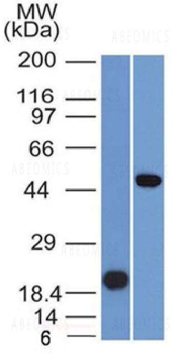
Fig. 1: Western Blot Analysis (A) Recombinant Protein(B) Raji cell lysate UsingPAX8 Mouse Monoclonal Antibody (PAX8/1491).
 Monoclonal Antibody(Clone: PAX8/1491)")
 Monoclonal Antibody(Clone: PAX8/1491)")
 Monoclonal Antibody(Clone: PAX8/1491)")
 Monoclonal Antibody(Clone: PAX8/1491)")
 Monoclonal Antibody(Clone: PAX8/1491)")
Roll over image to zoom in
Shipping Info:
For estimated delivery dates, please contact us at [email protected]
| Amount : | 100 µg |
| Isotype : | Mouse IgG2b, kappa |
| Content : | 200 µg/ml of Ab Purified from Bioreactor Concentrate by Protein A/G. Prepared in 10mM PBS with 0.05% BSA & 0.05% azide. Also available WITHOUT BSA & azide at 1.0mg/ml. |
| Storage condition : | Antibody with azide - store at 2 to 8°C. Antibody without azide - store at -20 to -80°C. Antibody is stable for 24 months. Non-hazardous. |
Recognizes a protein of 62kDa, identified as PAX8. It is a member of the paired box (PAX) family of transcription factors. This nuclear protein is involved in thyroid follicular cell development and expression of thyroid-specific genes. Mutations in this gene have been associated with thyroid dysgenesis, thyroid follicular carcinomas, and atypical thyroid adenomas. PAX-8 is expressed in the thyroid (and associated carcinomas), non-ciliated mucosal cells of the fallopian tubes, and simple ovarian inclusion cysts, but not normal ovarian surface epithelial cells. PAX-8 is expressed in a high percentage of ovarian serous, endometrioid, and clear cell carcinomas, but only rarely in primary ovarian mucinous adenocarcinomas. PAX-8 expression is reported in renal tubules as well as renal cell carcinoma, nephroblastoma, and seminoma. PAX-8 antibody may be used as an additional immunohistochemical marker for renal epithelial tumors.
Western Blot (1-2ug/ml); Immunohistochemistry (Formalin-fixed) (1-2ug/ml for 30 minutes at RT)(Staining of formalin-fixed tissues requires heating tissue sections in 10mM Tris with 1mM EDTA, pH 9.0, for 45 min at 95°C followed by cooling at RT for 20 minutes);
For Research Use Only. Not for use in diagnostic/therapeutics procedures.
|
There are currently no product reviews
|
")

 (Extracellular Domain) Monoclonal Antibody(Clone: TFRC/1818)")
 (JNJ-64304500)")
-Low Endotoxin")
")

")
")
 (Clone: 1E4) rabbit mAb PE conjugate")
 (Clone: ABM1C12)")


")
")
.png "ACE2/HEK293 Stable Cell Line")
")


(Clone : AE-4)")
")
 Monoclonal Antibody(Clone: 7.3)")


