

Monoclonal Antibody to GAP43 (Clone: 1E3) (Discontinued)
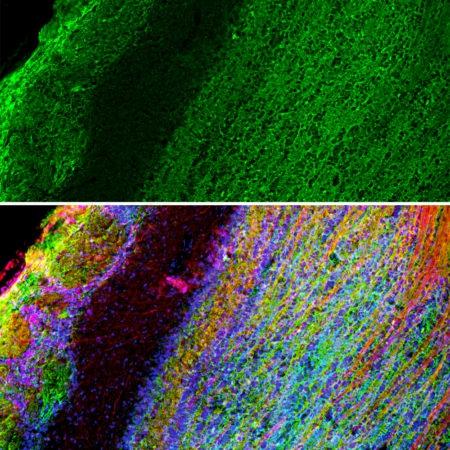
Figure 1: Mixed neuronal cultures stained with 34-1039 (green), MAP2, a rabbit antibody to microtubule associated protein 2 (MAP2, red) and DNA (blue). The GAP43 antibody stains the plasma membrane of neurons and is particularly concentrated in dendrites. Another image is here.
 (Discontinued)")
 (Discontinued)")
Roll over image to zoom in
Shipping Info:
For estimated delivery dates, please contact us at [email protected]
| Format : | Purified |
| Amount : | 100 µl |
| Isotype : | Mouse, IgG1 |
| Content : | Antibody is supplied as an aliquot of 1 mg/ml of affinity purified antibody or concentrated tissue culture supernatant. |
| Storage condition : | Store the antibody at 4°C; stable for 6 months. For long-term storage; store at -20°C. Avoid repeated freeze and thaw cycles. |
| Gene : | GAP43 |
| Gene ID : | 2596 |
| Uniprot ID : | P17677 |
| Alternative Name : | Axonal membrane protein GAP-43, Growth-associated protein 43, Neural phosphoprotein B-50, pp46 |
| Immunogen Information : | C-terminal peptide of rat and mouse GAP43, which is KEDPEADQEHA, with an N-terminal Cys added to allow chemical coupling to KLH carrier protein. |
Western blots: 1:10,000. IF/ICC and IHC: 1:1,000.
For Research Use Only. Not for use in diagnostic/therapeutics procedures.
| Subcellular location: | Cell membrane, Cell projection, Cell junction, Cell projection |
| Post transnational modification: | Palmitoylation by ARF6 is essential for plasma membrane association and axonal and dendritic filopodia induction. Deacylated by LYPLA2. |
| BioGrid: | 108867. 27 interactions. |
|
There are currently no product reviews
|
")

 (Clone: B9) rabbit mAb FITC conjugate")
(Discontinued)")
(Discontinued)")
")

")
 (Clone: G12) rabbit mAb SureLight488 conjugate")
 (Clone: ABM1C12)")


")
")
.png "ACE2/HEK293 Stable Cell Line")
")


")


